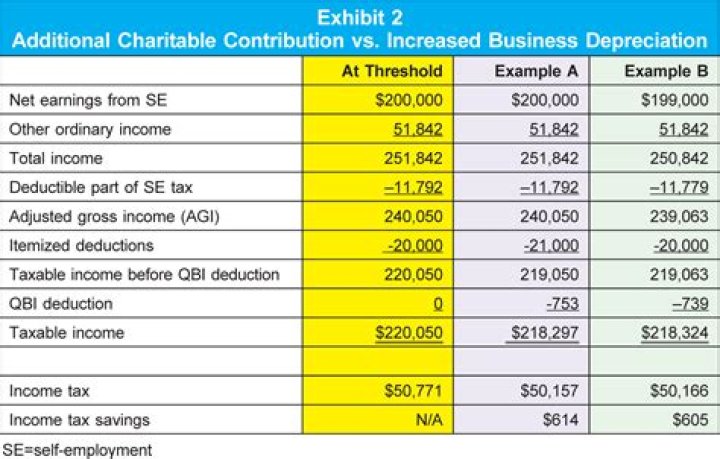
What is the LGN in the eye?
Optic nerve fibres from the eyes terminate at two bodies in the thalamus (a structure in the middle of the brain) known as the Lateral Geniculate Nuclei (or LGN for short). One LGN lies in the left hemisphere and the other lies in the right hemisphere.
Where is the LGN?
They wrap around the midbrain and cross the medial surface of the temporal lobe, and 80% of them then terminate in a synaptic relay called the lateral geniculate nucleus (LGN), located in the dorsal part of the thalamus. The LGN is thus the major target for each optic tract.
Why is the LGN important?
In addition to retinal afferents, the LGN receives input from multiple sources including striate cortex, the thalamic reticular nucleus (TRN), and the brainstem. The LGN therefore represents the first stage in the visual pathway at which cortical top-down feedback signals could affect information processing.
What does LGN stand for psychology?
The lateral geniculate nucleus (LGN) (lateral geniculate body or lateral geniculate complex), located in the thalamus, functions as a relay center for the visual pathway and receives the majority of its sensory input from the retina of the eye.
What does LGN stand for?
LGN
| Acronym | Definition |
|---|---|
| LGN | Look Good Naked |
| LGN | Logical Group Node (Sprint-ATM) |
| LGN | Logical Group Number |
| LGN | Linear Graph Notation |
What happens if the LGN is damaged?
In humans and other primates, visual information is transmitted from the retina to a part of the brain called the lateral geniculate nucleus (LGN), before reaching the primary visual cortex (V1). If the V1 is damaged, conscious vision is lost in the area of the visual field that corresponds to the damage.
Do the layers of the LGN separate left eye and right eye inputs?
Ipsilateral and contralateral layers Both the LGN in the right hemisphere and the LGN in the left hemisphere receive input from each eye. However, each LGN only receives information from one half of the visual field.
What cells are in the LGN?
The basis of the structure of the lateral geniculate nucleus is mostly in terms of its three distinct cell types: magnocellular (M), parvocellular (P), and koniocellular (K).
How does the LGN work?
The LGN brings retinotopic maps from both eyes into register to make it easy for cortex to combine inputs from the two eyes. Only 10% of inputs to LGN come from the retina. 90% are modulatory inputs from cortex and the brainstem.
What does LGM stand for in texting?
LGM
| Acronym | Definition |
|---|---|
| LGM | Let’s Get Married |
| LGM | Let’s Go Mets (New York Mets) |
| LGM | Little Green Men (Astronomical: first used as the designation for pulsars) |
| LGM | Lembaga Getah Malaysia (Malay: Malaysian Rubber Board) |
Where does the LGN receive information from the contralateral eye?
the eye on the opposite side (the contralateral eye) sends information to layers 1, 4 and 6. This description applies to the LGN of many primates, but not all. The sequence of layers receiving information from the ipsilateral and contralateral (opposite side of the head) eyes is different in the tarsier.
Where is the LGN located in the brain?
Although the retina sends axons to many subcortical nuclei, only the pathway from the retina to LGN to cortex is critical to visual awareness. The LGN is a distinctively layered structure and is located at the posterior lateral margin of the dorsal thalamus ( Fig. 29.1 ).
How is the map of the retina preserved in the LGN?
Magno (large) LGN cells receive inputs from (large) parasol ganglion cells. The retinal (hereafter called “retinotopic”) map is preserved. Axons from the retina preserve their order. There is an entire map of a visual hemi-field in each layer of the LGN. The maps are in register in each layer. LGN physiology
What is the role of the LGN in visual processing?
Given that visual signals are transformed in other visual areas it is puzzling that a similar transformation can not be identified in the LGN. The most reasonable explanation is that the main role of the LGN is to regulate the flow and strength of visual signals sent to V1.